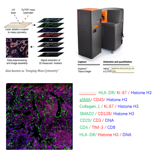
Imaging Mass Cytometry (IMC)
Mass Cytometry is an emerging multidimensional single-cell analysis technique that uses metal element-coupled antibodies combined with inductively coupled plasma mass spectrometry (ICP-MS) to detect labeled elements on single cells, avoiding the problems

Platform Introduction
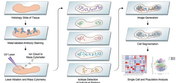
Mass Cytometry is an emerging multidimensional single cell analysis technique that uses metal element coupled antibodies in combination with inductively coupled plasma mass spectrometry (ICP-MS) to detect labeled elements on single cells, avoiding the problems of fluorescence crosstalk, autofluorescence and channel restriction of traditional flow-through methods. Antibodies labeled with metal elements recognize antigens on the cell surface or inside, and cells are sent one by one to the plasma torch for ionization, causing the release of labeled metal ions. The released metal ions are sent to a time-of-flight detection chamber for separation and detection, where the detector precisely records the arrival time of various ions, which in turn converts the precise amount of various metal labels in each cell. Finally, the data are analyzed using SPADE, PCA, viSNE and Gemstone methods.
(Expert Opin Drug Discov. 2019 Feb;14(2):115-125. )
Technical Advantages
The application of the metal labeling system has enabled IMC technology to greatly increase the number of markers that can be detected in a single experiment for a single section sample, making it possible to complete staining operations that require more than a dozen sections for immunohistochemistry (IHC) by traditional methods, now through a single section, effectively avoiding the discrepancies between data caused by sequential sections and the loss of samples during sequential staining, especially those It effectively avoids the discrepancy between data caused by consecutive sections and the loss of samples during consecutive staining, especially those precious and rare clinical samples accumulated in the clinic for many years, so that their value can be exploited to the maximum extent. At the same time, it is easier to discover new phenomena and results based on existing information and conclusions.
Applications
With the advantage of its multiparametric detection, IMC technology can identify an immune cell in tumor tissue and know to what subpopulation it belongs, as well as its location in the tissue and which cells interact with it in its surroundings. This simultaneous acquisition of localization and classification information is of great importance in the field of studying the tumor microenvironment, the fine mechanisms of tumor immunity, etc. It has been widely used in the fields of cell biology, molecular biology, immunology, hematology, drug screening and development, and clinical biomarker development for comprehensive and detailed research and analysis. 
Sampling Requirements
Slices need to be sealed with paraffin oil and stored at 4 degrees; wax block samples are stored at 4 degrees
Serial sections for easy targeting of target areas
Thickness less than or equal to 7μm
Tissue FFPE samples should be as fresh as possible, preferably made within six months, and paraffin blocks are preferred.